Visualizing intracellular nanostructures of living cells using
![](https://scx2.b-cdn.net/gfx/news/hires/2022/visualizing-intracellu.jpg)
![](https://wires.onlinelibrary.wiley.com/cms/asset/d12e44ef-afe3-461e-8562-d86843540ea4/wnan1486-toc-0001-m.jpg)
Methodologies and approaches for the analysis of cell–nanoparticle interactions - Ivask - 2018 - WIREs Nanomedicine and Nanobiotechnology - Wiley Online Library
![](https://pubs.rsc.org/image/article/2021/CC/d1cc02644e/d1cc02644e-s1_hi-res.gif)
Visualization of O 2 /ATP cross-talk in living cells with a smart fluorescent nanoprobe - Chemical Communications (RSC Publishing) DOI:10.1039/D1CC02644E
![](https://onlinelibrary.wiley.com/cms/asset/1c40a5c4-0120-4ef6-b622-c7dcba1f484c/ange202114267-fig-5001-m.jpg)
Supramolecular Self‐Assembly in Living Cells - Dergham - 2022 - Angewandte Chemie - Wiley Online Library
![](https://www.researchgate.net/publication/351047605/figure/fig5/AS:1015258923687967@1619067998757/Lanthanide-based-MOF-nanosheets-for-two-color-intracellular-ATP-imaging-in-living-cells.png)
Lanthanide-based MOF nanosheets for two-color intracellular ATP imaging
![](https://d1otjdv2bf0507.cloudfront.net/images/Article_Images/ImageForArticle_6008_16451766195089519.jpg)
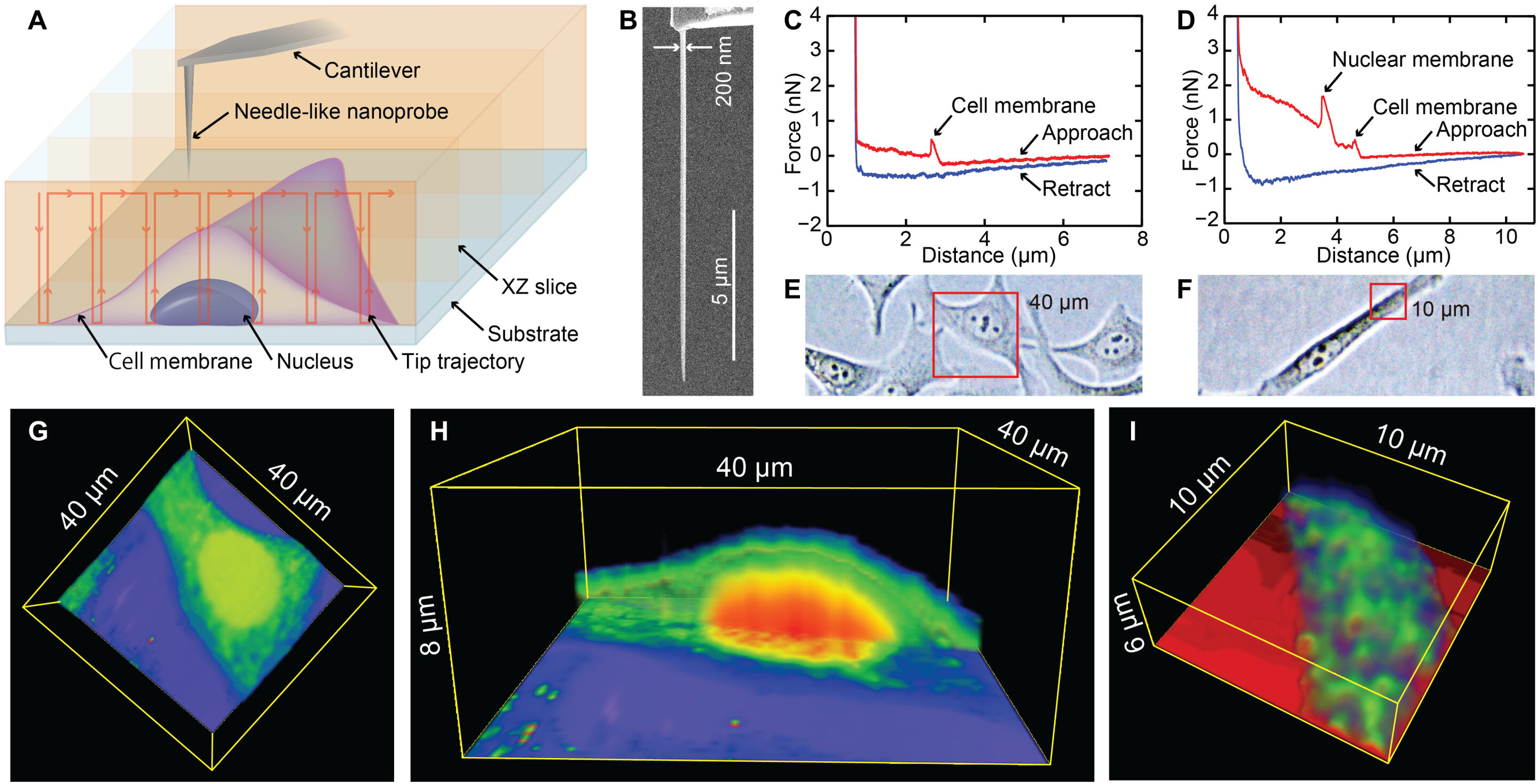
Nanoendoscopy-AFM: An Overview
![](https://www.researchgate.net/publication/347458822/figure/fig2/AS:986247006797825@1612151018660/Live-cell-holotomography-of-cellular-nanoparticles-PANC-1-SMAD4-2-6-cells-with_Q320.jpg)
Live-cell-holotomography of cellular nanoparticles. PANC-1 SMAD4 (2−6)
![](https://www.chinesechemsoc.org/cms/asset/59208d68-1eec-4a1c-b4bd-6002d33cf167/keyimage.jpg)
Light-Mediated Spatiotemporally Dynamic Assembly of DNA Nanostructures in Living Cells Regulates Autophagy
![](https://pubs.acs.org/cms/10.1021/nl102293b/asset/images/medium/nl-2010-02293b_0005.gif)
Visualizing Light-Triggered Release of Molecules Inside Living Cells
![](https://d3i71xaburhd42.cloudfront.net/293af99af9deafb6b91df29c089b1ab2a81f1626/13-Figure2-1.png)
PDF] Quantitative Visualization of Molecular Delivery and Uptake at Living Cells with Self-Referencing Scanning Ion Conductance Microscopy-Scanning Electrochemical Microscopy.