Histological section of the brain, case 2. Gitter cells (macrophages)
![](https://www.researchgate.net/publication/51450445/figure/fig3/AS:214044142247938@1428043508068/Histological-section-of-the-brain-case-2-Gitter-cells-macrophages-arrows-containing.png)
![](https://www.researchgate.net/profile/Jakob-Willesen/publication/51450445/figure/fig2/AS:214044142247937@1428043508043/Histological-section-of-the-brain-case-2-Haematoma-H-with-peripheral-fibroplasia_Q320.jpg)
Histological section of the brain, case 2. Gitter cells (macrophages)
![](https://www.frontiersin.org/files/MyHome%20Article%20Library/942967/942967_Thumb_400.jpg)
Frontiers Characterization of microglia/macrophage phenotypes in the spinal cord following intervertebral disc herniation
![](https://www.researchgate.net/profile/Jakob-Willesen/publication/51450445/figure/fig5/AS:214044142247940@1428043508113/Histological-section-of-the-lungs-case-3-Thrombosis-T-of-a-pulmonary-artery_Q320.jpg)
Histological section of the brain, case 2. Gitter cells (macrophages)
![](https://o.quizlet.com/APd-AOwnOvQA0DghUhGz.A.jpg)
Vascular Disorders of the CNS Flashcards
![](https://www.frontiersin.org/files/Articles/525240/fvets-07-00291-HTML/image_m/fvets-07-00291-g001.jpg)
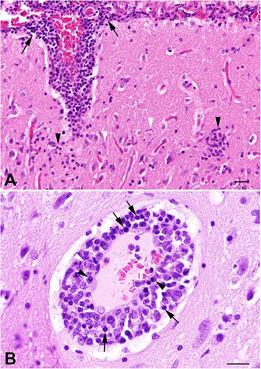
Frontiers Meningoencephalomyelitis of Unknown Origin in Cats: A Case Series Describing Clinical and Pathological Findings
![](https://ars.els-cdn.com/content/image/3-s2.0-B9780323449410000023-f002-002ab-9780323449410.jpg)
Normal Brain Histopathology - ScienceDirect
![](https://journals.sagepub.com/cms/10.1177/0300985812450719/asset/images/large/10.1177_0300985812450719-fig6.jpeg)
Nonlesions, Unusual Cell Types, and Postmortem Artifacts in the Central Nervous System of Domestic Animals - P. Wohlsein, U. Deschl, W. Baumgärtner, 2013
![](https://www.researchgate.net/publication/11949433/figure/fig2/AS:281738472378433@1444183094547/Histological-section-of-brain-from-a-sea-otter-containing-lesions-caused-by-and-stages-of.png)
Histological section of brain from a sea otter containing lesions
![](https://media.springernature.com/m685/springer-static/image/art%3A10.1186%2Fs12868-015-0226-7/MediaObjects/12868_2015_226_Fig6_HTML.gif)
Magnetic resonance imaging-based cerebral tissue classification reveals distinct spatiotemporal patterns of changes after stroke in non-human primates, BMC Neuroscience
![](https://media.springernature.com/lw685/springer-static/image/chp%3A10.1007%2F978-3-319-90495-5_126-1/MediaObjects/449709_0_En_126-1_Fig35_HTML.jpg)
General Principles of Ophthalmic Pathology
![](https://www.jneurosci.org/content/jneuro/30/25/8676/F1.large.jpg)
A Novel Population of Myeloid Cells Responding to Coxsackievirus Infection Assists in the Dissemination of Virus within the Neonatal CNS