A Custom Multiphoton Microscopy Platform for Live Imaging of Mouse Cornea and Conjunctiva
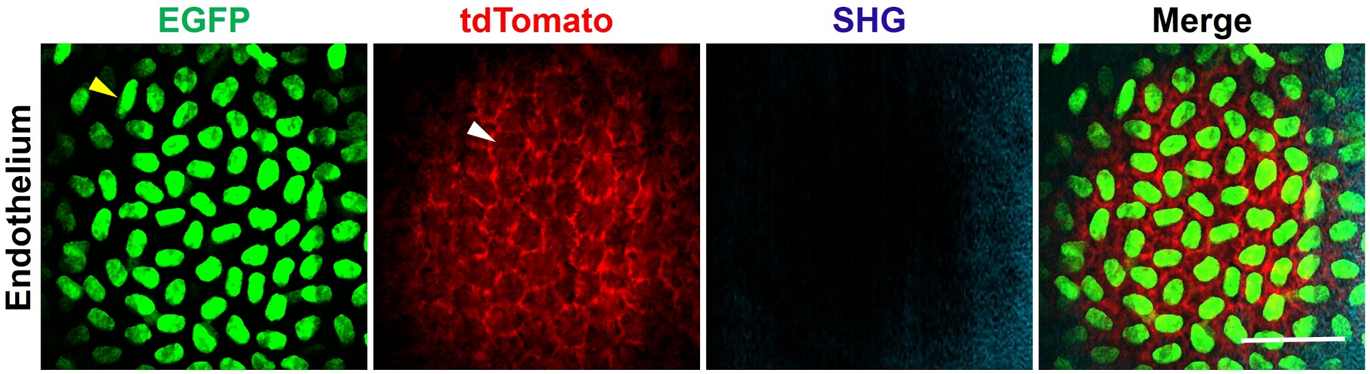
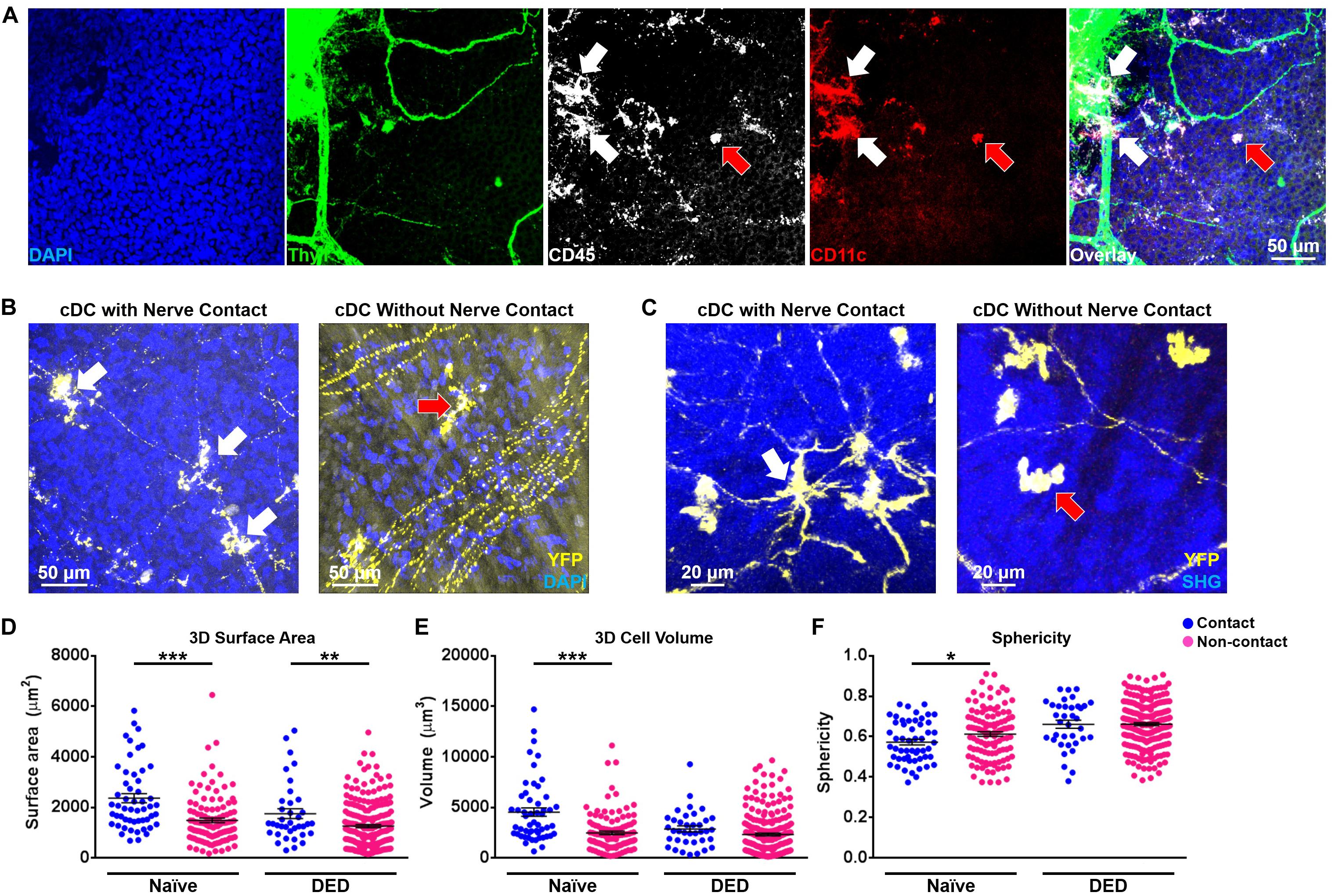
Frontiers Intravital Multiphoton Microscopy of the Ocular Surface: Alterations in Conventional Dendritic Cell Morphology and Kinetics in Dry Eye Disease

Two-photon live imaging of single corneal stem cells reveals compartmentalized organization of the limbal niche. - Abstract - Europe PMC

Posterior corneoscleral limbus: Architecture, stem cells, and clinical implications - ScienceDirect

Two-photon live imaging of single corneal stem cells reveals compartmentalized organization of the limbal niche - ScienceDirect

Autofluorescence multiphoton microscopy for visualization of tissue morphology and cellular dynamics in murine and human airways

Noninvasive two-photon microscopy imaging of mouse retina and retinal pigment epithelium through the pupil of the eye

A Custom Multiphoton Microscopy Platform for Live Imaging of Mouse Cornea and Conjunctiva

Two-photon live imaging of single corneal stem cells reveals compartmentalized organization of the limbal niche. - Abstract - Europe PMC

Bi-compartmentalized stem cell organization of the corneal limbal niche
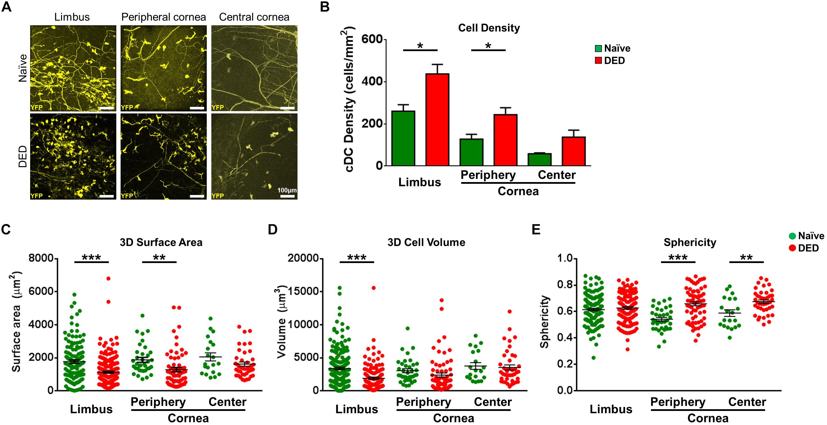
Frontiers Intravital Multiphoton Microscopy of the Ocular Surface: Alterations in Conventional Dendritic Cell Morphology and Kinetics in Dry Eye Disease

JCI - In vivo imaging of the human eye using a 2-photon-excited fluorescence scanning laser ophthalmoscope
Adaptive optics two-photon microscopy enables near-diffraction-limited and functional retinal imaging in vivo

Two-photon live imaging of single corneal stem cells reveals compartmentalized organization of the limbal niche - ScienceDirect

Two-photon microscopy of a Flt1 peptide–hyaluronate conjugate

Comparison of confocal microscopy and two-photon microscopy in mouse cornea in vivo.